Home Stretch | Repairing damage after a heart attack
A heart attack causes many heart cells to die and changes the structure of the heart muscle tissue, leaving the heart weaker and making it more difficult for it to pump blood around. TU/e PhD candidate Dylan Mostert investigated how to repair this tissue to regain normal heart function. This Friday, she will defend her dissertation at the Department of Biomedical Engineering.
The cardiac wall consists of three layers. The middle, thickest layer is the muscle layer – or myocardium – and its function is to generate the force needed to pump blood throughout the body. “What’s interesting about myocardial tissue is that it’s so nicely structured,” says Mostert. “It comprises a large number of layers – slices with a parallel organization of cells and tissue. Each of those slices is slightly rotated in relation to the next, creating a very efficient mechanism for pumping blood.”
Irreparable damage
When you suffer a heart attack, two major things happen, the PhD candidate explains. “Your myocardial wall no longer receives blood and oxygen, which results in the death of many beating heart cells. And in addition, the tissue loses that vital structure.” Heart cells can’t divide and become functional again, so unfortunately, the heart can’t regenerate itself. “If you sustain a wound to your skin, new tissue is produced. But any damage to the heart is severe, because it’s basically irreparable.”
After a heart attack, a thick scar tissue forms at the place where the heart cells died. “Imagine a little balloon, with one part of it being a lot stiffer than the rest,” she says. “Because that part is so stiff, the heart has to work harder and harder to pump blood with the same force.” The quality of life for these patients deteriorates dramatically – they become short of breath and their condition declines rapidly – their life expectancy also significantly decreases. “Because the heart has to work so hard all the time, this ultimately leads to heart failure,” Mostert says. Therefore, the main question at the heart of her research is: how can we restore the tissue structure after a heart attack and regain normal heart function?
Beating heart tissue
Heart cells cannot divide inside the body, but around the year 2007 it was discovered that it is possible to create heart cells in vitro (outside the body) from stem cells. “This led to extremely high expectations in terms of myocardial wall regeneration due to the huge potential of these cells!”, says Mostert. The idea was that you could create new heart cells and insert them back into the heart to repair the damaged tissue. But unfortunately, it turns out that is not how it works, she observes. “We’ve been trying to accomplish this for years, but it’s extremely complicated to restore heart function just by injecting new myocardial cells.”
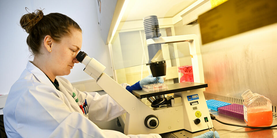
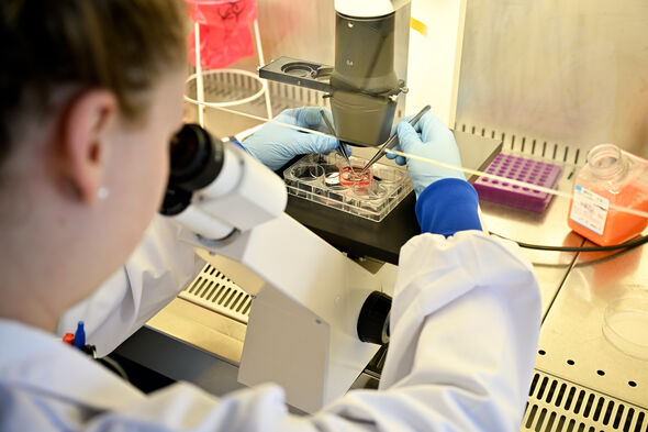
For that to work, it is essential to first restore the right structure in the tissue, she argues in her dissertation. To find out the best way to do this, she works in the lab with a tiny piece of beating heart tissue cultured from human stem cells. By leveraging the cells’ so-called mechanosensitivity (their sensitivity to mechanical stimuli), you can give the cells all kinds of stimuli and observe their responses. “This is how we discovered that we can control the structural organization of myocardial cells using a set of mechanical cues, such as stretching in the right direction,” she says.
A friend rather than an enemy
The heart muscle contains two important types of cells. Cardiomyocytes – or beating heart cells – are responsible for generating the force necessary to make the heart beat. The second type are cardiac fibroblasts. They create and organize the matrix and therefore play an important role in restoring the cells’ structure. If we can control these two types of cells and direct them in the right way, we can restore tissue organization,” Mostert asserts.
Mostert discovered that you can use fibroblasts, which are abundant in the heart muscle after a heart attack, for tissue reorganization. “Fibroblasts are often seen as the “bad cells”,” she says. During a heart attack, many cardiomyocytes die and are replaced by fibroblasts, which is not desirable. “But now we see that their presence is not necessarily a bad thing,” says Mostert. “If we direct them in the right way, they can actually help us repair the tissue. In that case, they can be a friend rather than an enemy.”
Young cells
Another important finding of her research is that the cell’s “age” affects its sensitivity. “Normally, you use a stem cell to cultivate a functional, beating heart cell in 21 to 40 days,” she explains. “But as it turns out, this heart cell is not very responsive to its environment.” She discovered that a cell on day 12 is much more sensitive and still susceptible to cues. “There have been so many attempts at inserting the cells into the heart, but they don’t connect to their environment and don’t become functional. If you were to place younger cells in there, it might work.”
She uses an analogy with a brick wall to illustrate her point. If the wall contains holes, it is difficult to fill them up with new bricks because those bricks are hard and need to fit exactly into those holes. Therefore, it is better to use clay because it is still completely malleable and easily adapts to its environment. “We’re now investigating whether these young cells can better adhere and integrate into the tissue and whether this can eventually lead to tissue organization and the restoration of function.”
Two celebrations
The PhD candidate has two major celebrations planned this year; in addition to receiving her PhD, she is getting married soon. “The last few years were sort of an in-between phase for me; it was a comfortable place where I didn’t have to make too many major life choices yet,” she says. Getting married and submitting her thesis feel like two huge steps toward adulthood for her. Both open new doors while closing others at the same time. “I do find that difficult at times, but mostly it’s very rewarding.”
PhD in the picture
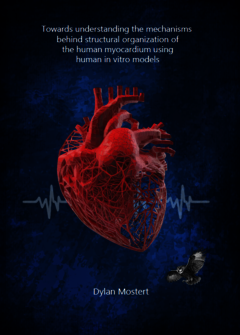
What is that on the cover of your dissertation?
“An image of the heart that clearly shows the structure of the tissue. In the background you can see the beating heart signal and in the corner is an owl – the companion of Athena, the Greek goddess of wisdom. ”
You’re at a birthday party. How do you explain your research in one sentence?
“I’m doing research on tissue repair after a heart attack.”
How do you blow off steam outside of your research?
“What works best for me is talking to friends about all sorts of other things, anything but my research. Other than that, I really enjoy cooking.”
How does your research contribute to society?
“We still have a long way to go, but I think I was successful in contributing a puzzle piece and providing many new avenues to explore, like investigating ways to restore organization and the maturity of cells used in stem cell therapy. And in the end, it’s all about the people, of course; the aim is to help all these different patients.”
What is your next step?
“I’m working on a postdoc right now and will finish in September. I’d like to stay in academia, for example as an assistant professor. My dream job is to be at the forefront of new research with a handful of people like me and really give it my all.”


Discussion