Customization for patients with abdominal aneurysm based on 3D ultrasound imaging
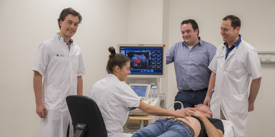
Researchers from TU/e and the Catharina Hospital in Eindhoven have succeeded in non-invasively measuring the elasticity and estimating the stresses in the human abdominal aorta using 3D ultrasound. Vascular surgeons can possibly use these metrics to better predict if and when an aneurysm, a dilation of the abdominal artery, should undergo a preventive operation to avoid a life-threatening internal hemorrhage. Unnecessary operations can also be avoided. Emiel van Disseldorp, PhD student at Biomedical Engineering, will be awarded his PhD for this research today, Thursday 10 January.
Aneurysms tend to occur in men over the age of sixty. To prevent them from rupturing, aneurysms with a diameter greater than 5.5 centimeters and fast-growing aneurysms are now operated on as standard. But in some patients the aneurysm ruptures anyway, while other aneurysms remain stable when the diameter is much greater. Therefore, doctors want to be able to predict for each patient who is at risk of the rupture, and on the other hand which patient is safe for the time being despite having a dilated aorta.
A balloon that is overinflated
The basis for the PhD research of Emiel van Disseldorp, led by Richard Lopata (Associate Professor at Biomedical Engineering) and vascular surgeon Marc van Sambeek, lies in biomechanics. An aneurysm does not rupture because of its size, but because the stresses in the vessel wall increase too much, just like with a balloon that is overinflated. Therefore, a method has been developed to measure the tension in the vessel wall and the stiffness of the aorta using ultrasound. The stiffness indicates to what extent a material can still deform in an elastic fashion, and changes in this can be an indicator for growth or even rupture of the aneurysm.
Since specialists want to be able to measure the stresses in and stiffness of the vessel wall quickly and regularly, this new method uses 3D ultrasound. This technique uses no radiation, is cheap and is easy to use. That is why 3D ultrasound is an ideal technique for this application. In this study, the technique for determining the vessel stiffness and the vessel stresses was further developed, resulting in the 'proof-of-principle'.
Clinical study
A clinical study has been set up with 320 patients and 30 healthy volunteers. Initial results show that vessel stiffness is an important value to measure the progression of the aneurysm. So far, in addition to the maximum diameter, the vessel stiffness and vessel pressure in 70 patients have been measured several times a year for more than 3.5 years.
The first results of this study show that a change in vessel stiffness is an important indicator in predicting growth and the possible rupture of the aneurysm. The researchers have already started undertaking further analysis of the research results to confirm these findings. Using this fully automated method, which was developed in-house, it can now easily be incorporated in large clinical studies.
Interested in reading more about this subject? You can find Emiel van Disseldorp's dissertation here.


Discussion