Home Stretch | Narrowing arteries seen in a new light
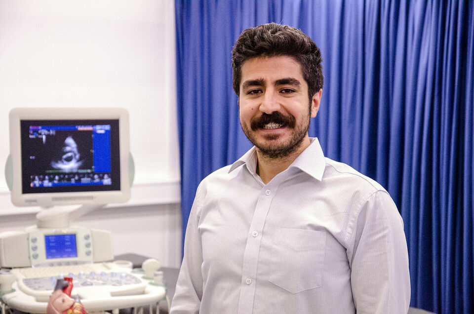
Thanks to a promising new method, cardiologists will in future be able to make a simple assessment of whether a high-risk operation on the carotid artery is necessary in cases of vascular constriction. Doctoral candidate Ümit Arabul worked in close cooperation with the Catharina Hospital to develop a handy photoacoustic scanner and improved the image processing of diseased carotid arteries.
Cardiovascular diseases are one of the major causes of death worldwide. These diseases are often a consequence of vascular constriction caused by ‘plaques’- deposits of cholesterol, other fat particles and inflammatory cells on the wall of the artery. Calcium can also accumulate in these plaques, causing blood vessels to calcify and further reducing the blood supply to essential organs like the heart and brain.
Moreover, as the plaque grows, tiny cracks in it may appear - now the plaque is ‘unstable’ - and it may eventually burst open. The plaque contents released in this way can cause tiny clots in the bloodstream, which when accompanied by a calcified carotid artery may lead to a stroke.
Matter of life and death
Sometimes, therefore, the early diagnosis of a narrowing of the arteries and an accurate assessment of the plaque composition are quite literally a matter of life and death, emphasizes Ümit Arabul. While doing his doctoral research at the Cardiovascular Biomechanics group (BMT) he developed a new method of visualizing atherosclerosis, as this narrowing is termed.
“Physicians currently assess plaques based on ultrasound images, familiar from pregnancy scans,” explains Arabul. “An excellent technique for studying the geometry of the atherosclerosis, but research is steadily revealing that it is the composition of the plaque, rather than its shape, that is the most important indicator of stability. How much fat and calcium are present, are minor bleeds already occurring in the mini-blood vessels in the plaque?”
“In order to study the plaque itself we decided to use photoacoustic imaging (PAI). Using a laser, a short pulse of light is directed into the body. Particles in the tissue at the study site absorb the light and convert it into heat. In so doing, the tissue expands rapidly and then collapses, and using a special probe this pressure change can be detected as ultrasound. This technique has existed for decades but our approach is new in terms of providing a different perspective in plaque imaging.”
Arabul’s research was part of a large European project - FULLPHASE - in which scientists studied whether photoacoustic imaging can be used to trace various diseases, ranging from cancer to rheumatoid arthritis, and including atherosclerosis.
“Early on in the project we developed a modified handheld photoacoustic scanner with built-in laser technology,” says Arabul. “By coupling this to an existing ultrasound device we were able to study in detail carotid arterial plaques - both their structure and composition - gathered from patient material. The images are promising: we can make concentrated regions of certain molecules in the plaque visible. Internal bleeds can also be seen clearly with this probe.”
Future
Owing to the highly positive results - and by using new image processing techniques, Arabul managed to rapidly improve the image quality - a new European study (CVENT) is focusing exclusively on the application of photoacoustic imaging in atherosclerosis. At TU/e three researchers recently picked up where Arabul's project left off. Thus he has no doubt that in the foreseeable future photoacoustic imaging will become part of clinical practice - and cardiologists, thanks to this technique, will be better able to estimate whether the state of the plaque necessitates a high-risk operation.
Arabul himself will not be seeing the follow-up at close hand. For four months now he has been working as a researcher at ASML, far removed from the biomedical field. Still, he hasn't left the world of images and sound behind him, he admits light-heartedly: “Same techniques, new applications”.


Discussion