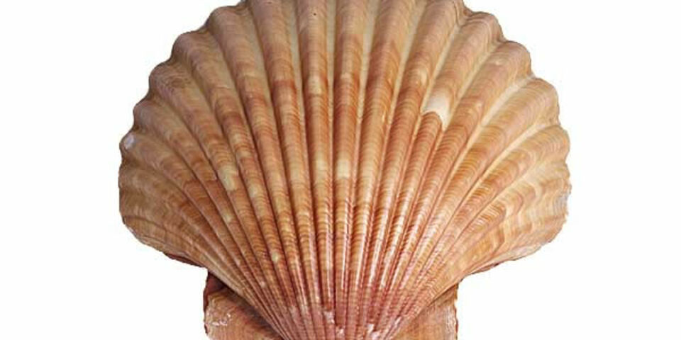
TU/e scientists see shells grow in real time
TU/e scientists, together with American co-workers, are able to observe the formation of shells in real time on a nanometer scale thanks to a new electron microscopy technique. This enabled them for the first time to see how pieces of polymer act as ‘ion sponges’ – thereby confirming a 30-year-old theory. The required ions are absorbed so that crystals are only formed at these specific locations. The researchers publish their result today in Nature Materials.
The findings not only throws a new light on biological crystal formation in nature, which is still not fully understood. The results also provide additional understanding of industrial crystal formation processes, which are used for example to increase efficiency in the production of ICs and solar cells.
Natural biomineralization, through which organisms and minerals grow, typically in shells and teeth, follows a process that technologists can only dream of. Our control of the present industrial crystallization processes is relatively primitive, compared with the perfection with which crystals grow and are ordered in nature. Therefore, scientists have for decades been trying desperately to understand and control these unique chemical processes in nature.
At the end of the 1980s, researchers in Israel presented a new theory about the first steps in the formation of shells, in which calcium and carbonate ions are present in solution shortly before they bond and crystallize into the well known, hard material: calcium carbonate. They believe that this crystallization process is initiated by surfaces of insoluble biopolymers that act as ‘ion sponge’: they attract calcium ions, so that they concentrate in these specific locations. The carbonate ions can then bond much more easily to the calcium, in a way that the calcium carbonate crystals only form at the specific locations of the sponges.
More than 30 years later, the technology is now advanced enough and researchers from TU/e, Pacific Northwest National Laboratory (PNNL) and Lawrence Berkeley National Laboratory led by Nico Sommerdijk (TU/e) and Jim de Yoreo (PNNL) have indeed seen how these ion sponges are formed. Using a new kind of electron microscopy technique, they have observed the formation of shells in a solution, in real time and on a nanometer scale. “This is a big difference from the earlier standard, in which you have to freeze samples. This means you could only study the individual steps in the process. Instead of studying ‘snapshots’ of those separate steps, we can now look at a ‘film’ of the complete process”, says Sommerdijk.
In the experiment, the polymer polystyrene sulfonate acts as an ‘ion sponge’. The solution to be studied was covered by thin, transparent membranes, forming a kind of ‘aquarium’. Using inlet valves, the researchers were able to allow ions of calcium and carbonate to flow into the experiment at will. In a first experiment without the polymer, few interesting effects were observed: the usual small crystals only formed occasionally. But with the addition of the polymer, it was clearly visible how the calcium was mainly adsorbed by the polymer, after which crystals formed primarily at these locations in the presence of the carbonate crystals. In addition the process was many times faster than without the polymer.
Crystallization is used in many technology applications, for example in nanotechnology or in the production of pharmaceuticals. “To get the crystallization process started just requires – in simple terms – the solution to be made less soluble”, Sommerdijk explains. “For example if I allow a warm solution of sugar to cool, the sugar will crystallize. But you never know where exactly the crystals will grow. With this kind of sponge, you can not only control where the crystals will grow, but also how the process will take place. This means you have much better control not only of their location, but also of their shape.”
Source: TU/e Press Team


Discussion