New predictive method can greatly reduce complications in hemodialysis
Approximately two million people around the world regularly undergo hemodialysis – blood purification outside the body – as a result of kidney failure. This usually involves creating a dialysis fistula – a connection between an artery and a vein – in the patient’s arm. However this causes complications in as many as 50% of patients, primarily related to changes in blood flow. Researcher Maarten Merkx of Eindhoven University of Technology (TU/e) has developed a system that automatically images the blood vessels in the arm and calculates the expected blood flow through the dialysis fistula. This makes it easy for physicians to find the best surgical strategy to insert the dialysis fistula with minimal chance of complications. Merkx gained his PhD on Monday 3 December for this research.
At present, physicians determine the location for the dialysis fistula using ultrasound images of the arm, a process similar to echography during pregnancy. The best location is different for each patient because of the individual differences in blood flow. The diameter of the blood vessels is measured manually from the images, but this is not a very accurate method . That makes it difficult to predict the blood flow through the dialysis fistula. If the blood flow through the fistula is too high it can result in heart failure and insufficient circulation through the hand, which can even cause loss of fingers. If the flow rate is too low it can lead to an inefficient dialysis process or other problems. These potential problems make more accurate prediction of the blood flow highly desirable.
Together with researchers from Maastricht University Medical Centre (MUMC) and other institutes, Maarten Merkx has developed a system that automatically calculates the blood flow in the blood vessels of the arm, based on an MRI scan. His research shows that this system allows good prediction of the blood-flow effects, caused by the insertion of the dialysis fistula, in virtually all patients. An important part of Merkx’s system is the automatic determination of the diameter of the blood vessels. This has proved to be much more precise than the present manual method: his system has a maximum error of 3%, compared with at least 20% for the manual method.
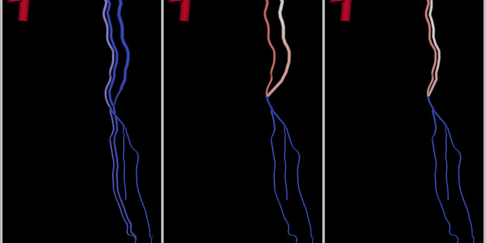
The system developed by Merkx converts the MRI images into a kind of ‘wiring diagram’ of the blood vessel system in the arm. A physician can see the calculated flow rate at any point in the network, which enables the best place to insert a dialysis fistula to be located. A particularly important aspect is the choice between the lower or upper arm.
The introduction of Merkx’s technique can lead to a significant reduction in the number of complications arising in hemodialysis. Merkx expects that it will take at least five years before the technique is widely available. The first step is a clinical study which is now planned among a large group of kidney patients for further testing and development of the technique. Despite the use of MRI, which is relatively costly, Merkx believes that his technique will lead to lower costs due to the reduction in the number of complications.
Maarten Merkx gained his PhD on Monday 3 December 2012 for his doctoral thesis ‘Application of image-based computational modeling to vascular access surgery’. His work forms part of the ARCH project (http://www.vph-arch.eu/), which is carried out by a consortium of companies and knowledge institutes with financial support from the European Union (FP7). At TU/e the work is a result of the synergies between the Biomedical Image Analysis group led by prof. Bart ter Haar Romeny and the Cardiovascular Biomechanics group of prof. Frans van de Vosse, who also acted as Merkx’s lead supervisor.


Discussion