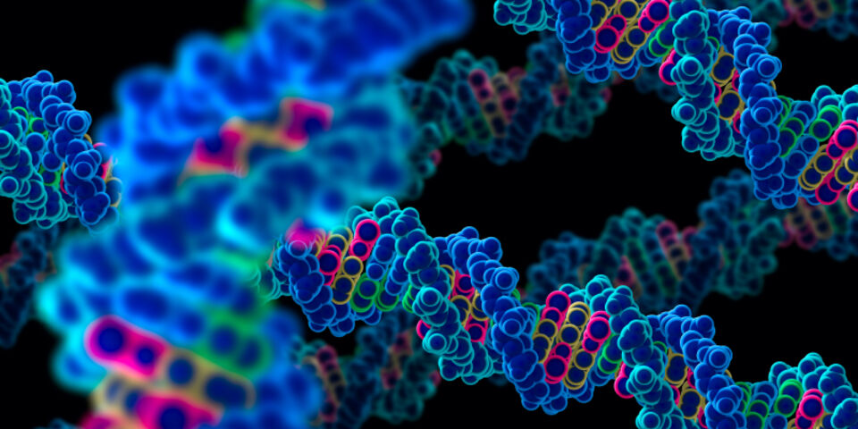
How do you represent DNA directly?
Thanks in part to TV series like CSI, the audience has been introduced to DNA and DNA analysis techniques. DNA is often pictured as the twisted ladder-like structure we all know, or as colored peak- the DNA sequence. Visualizing DNA as a molecule is still a very specialized area of expertise, and that’s why the genetic material is often shown indirectly. Last week, several media reported about an Italian research group that had managed to represent the strands of DNA for the very first time. And there it was indeed: a double helix. Is it very special to picture our genetic material this way, and why is it that DNA doesn’t like its picture taken?
“The article in question that appeared in Nano Letters featured some wonderful images, no doubt about that. Still, the first thing I thought, was: I’ve seen this before”, says dr. Nico Sommerdijk, associate professor of Materials and Interface at the Department of Chemical Engineering & Chemistry, and involved in the founding of the TU/e Soft Matter CryoTEM Research Unit. “It turns out directly visualizing DNA is not new. Already in 1995 did researchers show a double helix in watery surroundings with the help of Atomic Force Microscopy (AFM). With AFM, a microscopic needle scans the surface, enabling a very high resolution of the nanometers. A DNA molecule measures approximately 3 nanometers, so AFM has no trouble showing it. The media stating that DNA has never been pictured directly before is therefore just not right.”
“What is new, however, is the fact it’s been pictured without any background, so there’s no interference. By stretching groups of DNA helixes around two ‘pickets’ and drying those, a clothesline of sorts is formed in between the pickets: a floating group of DNA strands, which can be photographed beautifully by means of an electron microscope. The calculations they’re doing on the strands might be less trustworthy. After all, who says the drying process doesn’t create tensions that influence mutual distances?”
“Since DNA is so small, you’ll need high-resolution techniques such as electron microscopy (EM). At the same time, anyone prefers to study the DNA molecule in its natural -watery- surroundings, and DNA is very vulnerable at that. All of that doesn’t help. High-resolution EM methods used for material research are unsuitable for DNA, because it will literally be scorched by the strong electron beam. And EM methods from biology are too limited to properly picture DNA."
"Over the past years, the methods from these two areas have been merging -one of the strong suits of our own CryoTEM Unit- and that opens up new possibilities. For example, we’re currently working on the development of liquid-cell-TEM, in which molecules and nanoparticles in a tiny liquid-cell can be viewed in high resolution. For DNA, that means we can meticulously study the replication mechanisms, as well as find out if and where the genetic code fails. The ultimate dream is to be able to see the genetic letters individually. For now, that would mean fundamental research, although sequencing DNA should be feasible as well. But as you can tell from CSI, there are much faster and easier ways to do that.”


Discussion