
- Home Stretch
- 25/03/2025

Home Stretch | Depression under the scan
Greater understanding of how depression progresses thanks to brain scan
Depression affects millions of people worldwide, but much is still unknown about this illness and treatments don’t always work. PhD candidate Jesper Pilmeyer examined patients’ brains using MRI technology. The patterns he discovered in the brain scans can help make a more accurate diagnosis and predict how the illness will progress.
“Although depression affects one in six people, we still know extremely little about this mental illness,” says Jesper Pilmeyer. There are several theories about the causes of depression. “We know that a hereditary factor comes into play and that it may be related to a disturbance in the uptake of serotonin – the happiness hormone. A shortage of this can be remedied with antidepressants, but this only works in some of the patients.”
Depression is being looked at from different angles in hopes of gaining more insight into the illness. Pilmeyer’s research focused specifically on abnormalities in patients’ brains. By thoroughly analyzing dozens of MRI scans, he aimed to discover patterns that would allow for better predictions of how the illness progresses.
Biomarkers
The PhD candidate, with a background in Biomedical Engineering, began his research at the Department of Electrical Engineering. “In our Signal Processing Systems research group, we focus on analyzing and interpreting signals and images,” he says. “I’m in a subgroup that deals with biomedical diagnostics.” This includes magnetic resonance imaging (MRI), a technique that uses strong magnets and radio waves to create detailed images of the inside of the body without harmful X-rays.
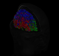
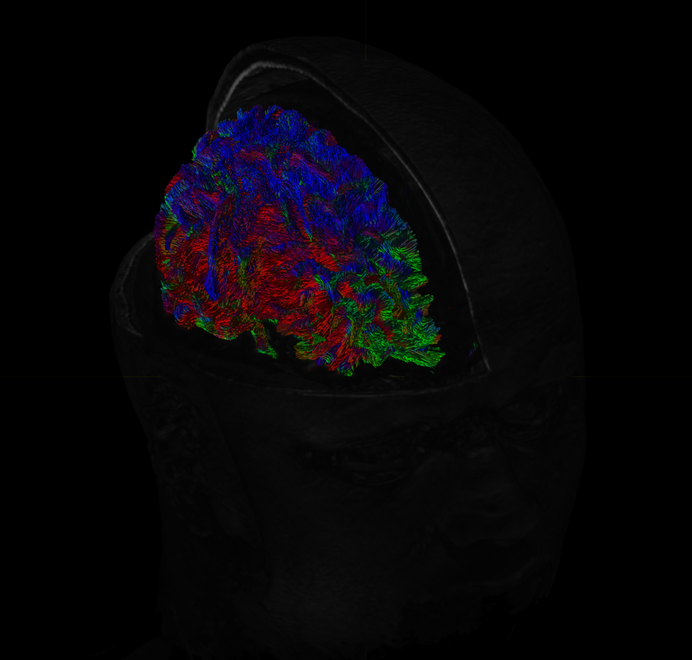
“With MRI, you can measure different biological features. For example, a functional MRI (fMRI) focuses on brain activity by indirectly measuring blood changes in different brain regions,” Pilmeyer explains. “But you can also analyze the physical structure of tissues and see how they’re connected to each other, as they form different networks together.”
Using these measurable biological characteristics – also known as biomarkers – we could diagnose depression more objectively and accurately. “This is currently done only on the basis of subjective symptoms such as gloom, fatigue, or anxiety, which can vary from patient to patient,” he explains. But more importantly, analyzing these biomarkers could help us predict how the illness will develop in different patients.
Under the scan
Pilmeyer’s primary research focus was on measuring brain activity. “You have different brain networks with a specific function. For example, there’s a visual network that’s activated when something moves in your field of view,” he explains. “We started looking at how these functional brain networks communicate with each other and whether we could discover interesting patterns in them.”
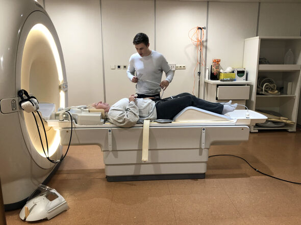
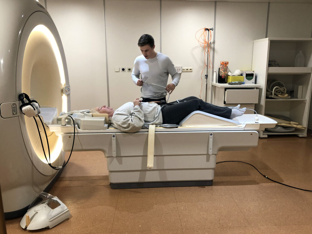
To examine data from real patients, he set up his own clinical study in collaboration with the Kempenhaeghe clinic, Philips Research, and GGzE. “Completely from scratch,” he emphasizes. “In the end we had about thirty patients participating, as well as an equally large, healthy control group.” On day one, scans were taken in both groups to compare them with each other. “The observed differences can help in understanding the pathology of depression,” he explains. In the patients with depression, new scans were taken every three months to study how the illness progresses. “This allowed us to see if we could use MRI to predict whether symptoms get better or worse over time.”
Pilmeyer was present himself when the scans were taken, which meant he personally interacted with the patients who participated in the study. “Afterwards they would often come up to me for a chat, asking all kinds of questions out of curiosity,” he says. The researcher found it valuable to observe everything up close like this. “Not only does it give you a better understanding of the technology involved in making the scans, but the direct contact with the patients also made the goal of the study – to help patients – very concrete and therefore played a key part in my motivation throughout the process.”
Dynamic communication patterns
The analyses of the scans provided many interesting insights. “Many existing studies examined how two networks work synchronously, or in other words, how they’re activated simultaneously,” he explains. “However, we found that it’s also possible for the activation of one network to deactivate the other, so you see an anti-correlation. In addition, the activation of one network may cause another network to become active somewhat later.”
These are all examples of dynamic communication patterns that can provide valuable information. “We saw that these patterns predicted how the illness would develop much better than the static patterns that are usually studied,” Pilmeyer says.
A more complete picture
Although the findings of his research need further validation in a larger study with more patients, they’re quite promising already. “The patterns discovered in the MRI scans can hopefully reveal more and more about this illness,” he says.
Incidentally, there are different subgroups of patients with depression; not everyone is the same, the researcher emphasizes. “There are studies that show that there are patients with brain activity abnormalities in whom certain medicines work better than in others. We hope that in the future, based on the measurements in the brain, we’ll be able to choose an appropriate treatment for each patient.”
It will probably never be possible to make diagnoses, predictions, and treatments based solely on MRI, Pilmeyer concedes. “We’ll always have to take other factors, such as heredity, into account.” Nevertheless, brain scans can provide valuable information that helps us get a more complete picture of the illness. “This could eventually contribute to more effective treatments, making a difference for millions of patients,” he concludes.
PhD in the picture
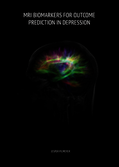
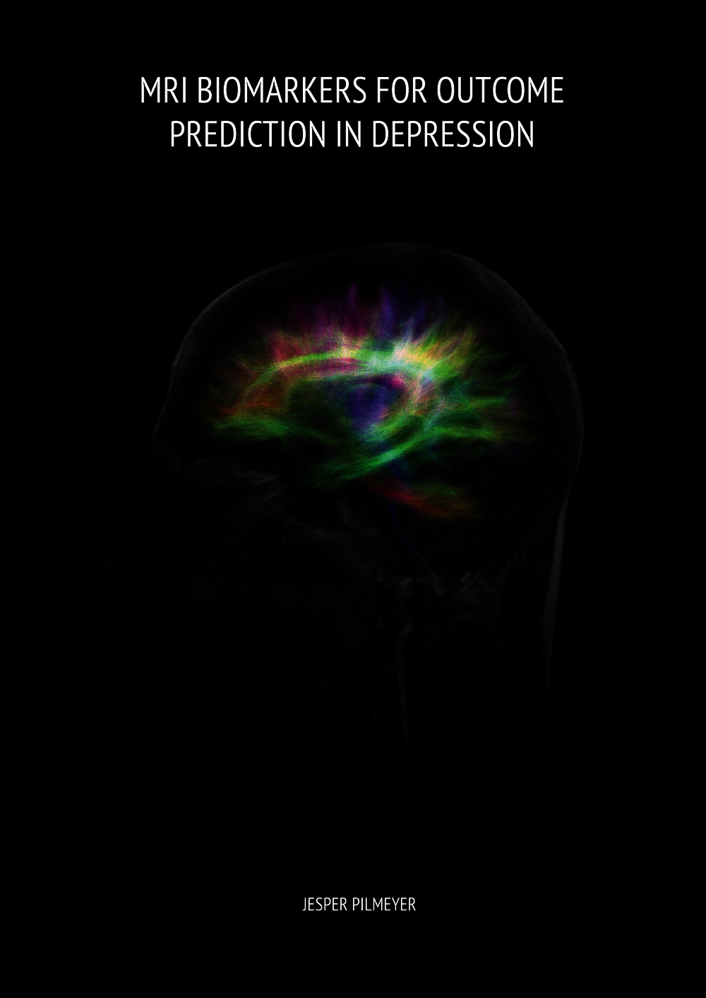
What is that on the cover of your dissertation?
“An MRI scan of my own brain. We had to optimize the MRI settings and I often was the test subject for this, so I think I was in there about a dozen times. This also allowed me to see if the quality of my data was good enough.”
And how does it feel under the MRI machine? “A scan takes a little over an hour. You lie still and aren’t allowed to do anything, so I often fell asleep in there,” he says, laughing.
You’re at a birthday party. How do you explain your research in one sentence?
“I study MRI scans of the brains of people suffering from depression to predict how the illness will progress.”
How do you blow off steam outside of your research?
“I like to play soccer with my teammates from the building where I live. Afterwards, I’m completely destroyed. I also organize board game nights with different groups of friends, which I love to do!”
What tip would you have liked to receive as a beginning PhD candidate?
“Don’t worry too much about the number of publications. You really don’t have to lose any sleep over the first one taking a while to appear. With me, there was nothing for a long time, and almost all my publications came right at the end. It always works out.”
What is your next step?
“I’m still applying for jobs. I hope to find something in industry or in a hospital – something that’s practice-based in any case. I would like to continue working on data-driven research within biomedical technology and signal processing. That could be MRI, but also CT scans or ultrasound. There are so many things I find interesting.”
Discussion