- Research
- 8 min
- 01/10/2024

Every breast is unique
October Breast Cancer Awareness Month: AI optimizes radiation planning for breast cancer
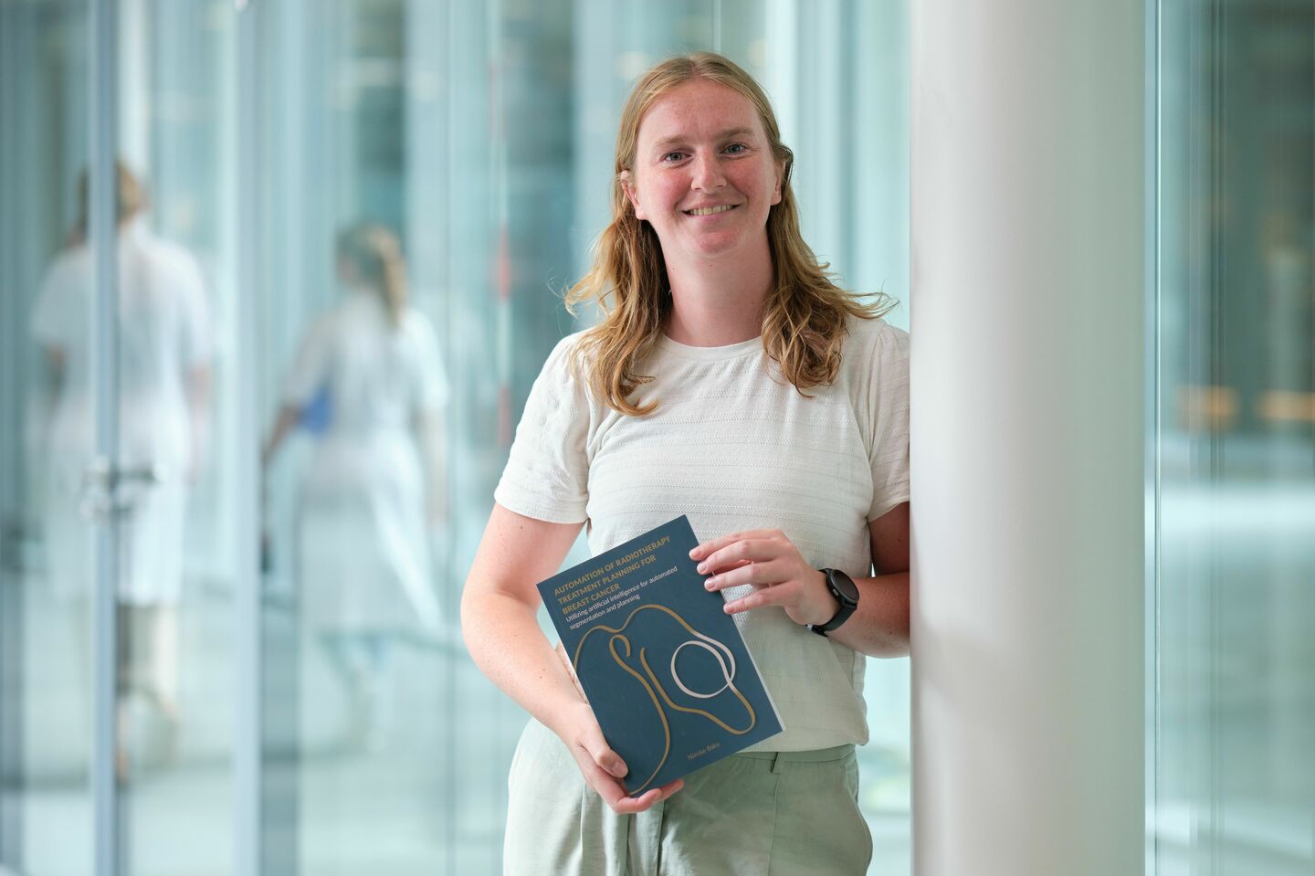
Every year, the month of October is globally dedicated to breast cancer, the most prevalent form of cancer among women. There is a lot of research focused on improving breast cancer treatments. One of these researchers is TU/e’s Nienke Bakx. During her doctoral period, she developed an AI model that saves radiation technicians and radiotherapists a lot of time when preparing a radiation treatment plan.
Last year, approximately 18,000 people were diagnosed with breast cancer in the Netherlands. More than half of these breast cancer patients are likely to see a radiotherapist for radiation treatment. Radiation treatment ensures that any remaining cancer cells are destroyed following surgery so as to minimize the chance of breast cancer recurrence.
Time-consuming preparations
However, before a breast cancer patient can start the treatment under the radiation machine, a lot of time goes into the preparations, Nienke Bakx knows. She has worked for quite some time as an AI expert at the Catharina Hospital in Eindhoven. She first started her graduate internship there, and then she stayed on to conduct research for her EngD Qualified Medical Engineering program. Her research ultimately yielded such a wealth of results that she was recently able to defend her dissertation. “Is it an EngD with a strong focus on research, or just a very practical PhD?” the brand-new doctor wonders with a smile. Either way, the Radiotherapy Department is now saving a lot of time thanks to her research.
One question is central to all of Bakx’s studies: how can we utilize artificial intelligence in planning radiation treatments for breast cancer patients? Bakx: “Such a treatment plan requires great precision and is unique for each patient. First, you must segment the organs very accurately – the tumor area should receive as much radiation as possible and the surrounding organs, such as the heart and lungs, should be protected from the harmful radiation as much as possible. Then, it’s time for calculations: how much and how many times should radiation be administered, and from which angle? When I started working at the Catharina Hospital five years ago, radiation technicians and radiotherapists would perform this entire process manually for each patient. After all, every breast is unique. It was a very time-consuming task.”
Complex breast
Bakx examined whether it was possible to optimize the radiation treatment plan using automatic calculations. “In most cases, patients have already undergone surgery to remove tumor tissue from the breast. I started working on a model that can determine the required dose for each area after segmentation of the surrounding organs.” Collecting enough suitable data in particular took a long time. That is something many AI researchers struggle with, Bakx explains. “You use older patient data, which all differ from each other, due to different protocols or equipment. And I specifically wanted to create a model for the more complex left breast, for which you need to take the heart and lungs into account during radiation treatment. Together with two experienced radiation technicians – who know exactly what they’re looking at – we went through all the data. And since breast cancer is unfortunately so common, there was a mountain of data available. A bittersweet silver lining. With all the usable previously created plans, I could train my computer model to ultimately create a radiation plan by itself.”
The initial results proved fruitful; and the Radiotherapy Department has been using AI-generated radiation treatment plans for several years now. After completing her EngD program, Bakx decided to continue her research. She was eager to take the next step: can we also have a computer perform the organ segmentation process more efficiently and reliably? “I started feeding a new model images of segmented organs. In a clinical pilot study, we compared manual and automatically generated segmentations. We were pleasantly surprised: 90% of the AI images were usable without further adjustments, and even when adjustments were required, they resulted in time savings. This provides a great starting point for the radiation technician. The computer model is not sacred, after all; the human eye remains essential.”
Future-proof healthcare
How is the use of AI in the treatment process viewed in the workplace? “When I started working here, the application of AI in healthcare was much less common than it is now; developments are advancing at breakneck speed. Some doctors are still somewhat skeptical. Through open communication, sharing positive results and providing training, we try to bring everyone on board.” There is plenty of room for discussion, Bakx emphasizes. “What works well in the plans and what could be improved? We want to support the doctors and technicians in their work; they are the ones who will actually be working with it. By automating certain processes, they get to apply their expertise elsewhere.”
The close collaboration with software provider RaySearch Laboratories, a Swedish company connected with major cancer institutes worldwide in order to improve treatment methods, also helps in establishing a more efficient working method. Bakx: “Those direct lines of communication are beneficial for making data and the necessary infrastructure available. In just a few years, we have transformed data into a usable model. This helps to improve care for breast cancer patients at the Catharina Hospital, and with our AI radiation planning we are among the forerunners worldwide. Through new research, we hope to make the AI model future-proof so that it can also be applied to other types of cancer. Because the ultimate goal is to provide optimal care for every patient.”
Discussion